baseline vaginal ultrasound
On the plus side you are not required to have a full bladder for this ultrasound. Your doctor will insert a speculum just like for a Pap test and cleanse the cervix with a betadyne soap solution.
![]()
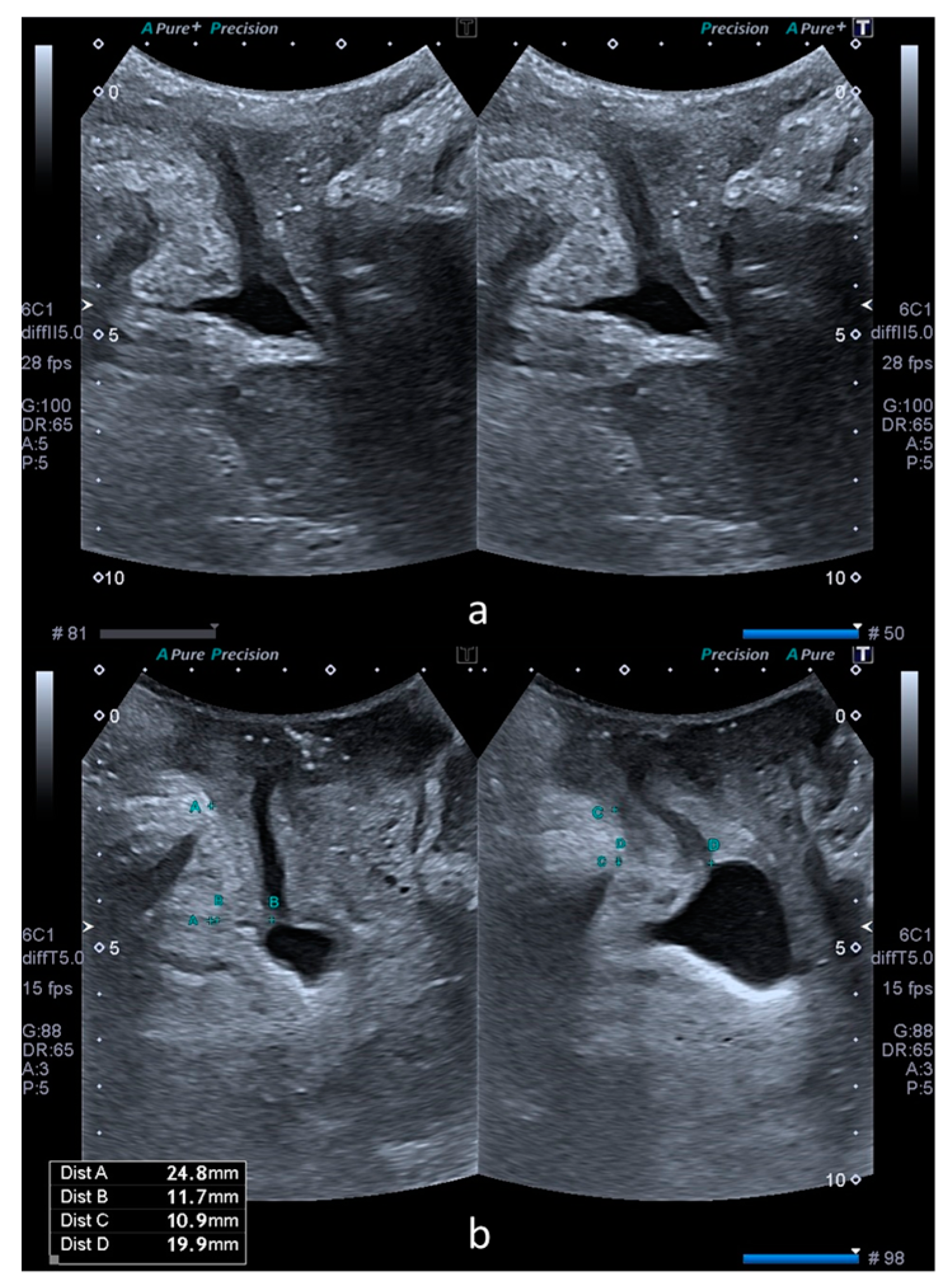
Transverse Ts And Longitudinal Ls Transvaginal Ultrasound Images Of Download Scientific Diagram
12 Sometimes a stubborn corpus luteum cyst sticks around even after your period starts.
. Unlike men who produce sperm on an ongoing basis females are born with a lifetime supply of eggs in their ovaries. There are two phases to a baseline ultrasound. Point-of-care ultrasound versus radiology department pelvic ultrasound on emergency department length of stay World Journal of Emergency Medicine 73 178.
The baseline ultrasound for an intrauterine insemination IUI is the same as a baseline scan for an IVF cycle. Ultrasonography of uterine leiomyomas Menopausal Review 164 113-117. To start we will apply a gel to the patients abdomen and run a probe along their midsection.
The mean creatinine in patients with vesicovaginal fistulas VVF was 060 ngml versus patients with uretero-vaginal fistulas UVF 079 ngml P 0012. What to expect in your baseline ultrasound. Transvaginal ultrasound scan An ultrasound scan is a procedure that uses high frequency sound waves to create a picture of a part of the inside of your body.
The ultrasound scanner has a probe that gives off sound waves. This article discusses the scan and the conditions that it can pick up on as well as some general points about fertility and health. In some cases an endometrial sample or biopsy may be performed.
If you are undergoing infertility treatment you will have a baseline ultrasound where we check to confirm that your ovaries have not formed any unexpected cysts that may disrupt your treatment. Ultrasound monitoring may begin on day 3 of the cycle to assess a baseline size as well as exclude if any cyst remains from previous hyperstimulation or otherwise. This is performed to view the endometrium uterus lining including its thickness and any associated ovarian abnormality.
This includes the uterus fallopian tubes. This is known as your baseline ultrasound. We also look at the endometrial lining thickness to make sure it.
All abnormal findings are measured and characterized such as fibroids uterine malformations hydrosalpinges and ovarian cysts. A transvaginal ultrasound is an internal scan of the female reproductive organs. Similarly vaginal pressure readings at.
A pelvic ultrasound is a noninvasive diagnostic exam that produces images that are used to assess organs and structures within the female pelvis. Transvaginal ultrasound is preferred and usually mandatory modality for monitoring follicles. The doctor will insert the transducer into the vagina after you empty your bladder.
A transvaginal ultrasound also called an endovaginal ultrasound is a type of pelvic ultrasound used by doctors to examine female reproductive organs. A transvaginal ultrasound showing the heartbeat of a fetus early in a pregnancy. Ultrasound remains one of the most routinely performed medical investigations and a mainstay of clinical decision-making rests upon the obtained results.
The lines at the bottom are called M Mode which is how imaging specialists detect and document heart motion. An adnexal mass in the pelvis shown with traditional ultrasound. The sound waves bounce off the organs inside your body and the probe picks them up.
An abdominal ultrasound and a vaginal ultrasound. Woźniak A Woźniak S. The doctor will usually perform a baseline transvaginal first.
This isnt dangerous and will usually go away without intervention. Although the bladder does not need to be full to perform this portion of the ultrasound it also should not be empty. After the ultrasound is complete the vaginal probe will be removed.
An adnexal mass shown more clearly with a transvaginal ultrasound. Due to the relative proximity of the pelvic organs to the abdominal surface and easy access via the vaginal route gynaecological scanning should be in the armamentarium of every gynaecologist. A baseline pelvic ultrasound is an internal vaginal ultrasound which is recommended for most patients as a part of their initial evaluation before starting treatments.
Baseline or screening ultrasound to assess the pelvic anatomy of the uterus including uterine lining and bilateral ovaries. It involves inserting a small ultrasound probe called a transducer into the vagina to produce incredibly detailed. A pelvic ultrasound allows quick visualization of the female pelvic organs and structures including the uterus cervix vagina fallopian tubes and ovaries.
Unlike IVF an IUI cycle typically use oral ovulation induction medications like Clomid or. Baseline ultrasound Around the time of your expected period we will perform a transvaginal ultrasound scan to examine your ovaries. The probe looks a bit like a microphone.
One of the baseline tests for female fertility and reproductive health is the General Pelvic Ultrasound Scan a quick and easy way to examine the structures essential for healthy female reproductive function. Their contents often show internal echoes because of the mucin content or due to infection. Doctors perform transvaginal ultrasound very much like a gynecologic exam.
When a grade 3 or more hydronephrosis is absent on renal ultrasound the negative predictive value of the presence of UVF is 933 95 confidence interval CI 886-962 with a specificity of 972 95 CI 903. The purpose is to check that there are no unusual cysts on the ovaries before starting the fertility drugs. However it is more invasive as it requires the ultrasound transducer to be inserted into the vagina rather than just rested on the abdomen.
This procedure is used to ensure your ovaries are not producing eggs at the moment are suppressed. If you are or have been on Tamoxifen Armidex Anastrozole or Femara it is important that you get a baseline ultrasound of your uterus and then follow up with a second ultrasound two years later. 64 and 65 These cysts are seen located posterolateral to the vaginal introitus medial to labia minora.
This kind of ultrasound provides a clear evaluation of the uterus and ovaries allowing for the diagnosis of uterine masses or ovarian cysts. The ultrasound is still done on cycle day 2-3 of your menstrual cycle and will help determine if you can start your IUI cycle that day. It also measures your serum estradiol level.
The ovarian reserve reflects her fertility potential. Transvaginal Ultrasound Scan This type of scan produces a higher quality image than the transabdominal ultrasound. They are seen as unilocular well-defined cysts usually 14 cm in size.
In some cases women may develop cysts. Before I started these drugs my first ultrasound 2 years ago showed my uterus to be in perfect condition. Iui infertility vaginalultrasoundWatch as I discuss what the baseline ultrasound process was like what the doctor is looking for and what to expect next.
Its important to count the number of existing follicles document twothree dimensions. Ultrasound Features of Bartholin Gland Cysts Figs. The Basal Antral Follicle Count test is a transvaginal ultrasound study that measures a womans ovarian reserve or her remaining egg supply.
Although improvements were seen in VAS scores for dysmenorrhea nonmenstrual pelvic pain and dyspareunia there were no significant differences in ultrasound biometry at either rest Valsalva or on contraction when comparing postinjection measurements at 4 12 and 26 weeks with pre-injection baseline. Ultrasound uses a transducer that sends out. A thin catheter tube will then be inserted into your cervical canal to enter the uterine cavity.
From the case rID.

The Evolving Role Of Saline Infusion Sonography Sis In Infertility European Journal Of Obstetrics And Gynecology And Reproductive Biology
![]()
Laparoscope And Transvaginal Ultrasound Images During Ablation Download Scientific Diagram
2
![]()
Transvaginal Ultrasound Of Uterine Myoma And Gestational Sac Gs At 8 Download Scientific Diagram
![]()
Transvaginal Ultrasound Of The Rectosigmoid With Signs Of Infiltration Download Scientific Diagram

Second Trimester Transvaginal Ultrasound Measurement Of Cervical Length For Prediction Of Preterm Birth A Blinded Prospective Multicentre Diagnostic Accuracy Study Kuusela 2021 Bjog An International Journal Of Obstetrics Amp Gynaecology

Chapter 14 Sonographic Assessment Of Early Pregnancy Obgyn Key
![]()
Laparoscope And Transvaginal Ultrasound Images During Ablation Download Scientific Diagram

Pelvic Ultrasound Showing The Large Complex Ovarian Cyst With Layered Download Scientific Diagram
Medicina Free Full Text Transperineal Ultrasound Assessment Of A Cystocele S Impact On The Bladder Neck Mobility In Women With Stress Urinary Incontinence Html
![]()
Sagittal View Of The Uterus On Trans Vaginal Ultrasound A Bulky Download Scientific Diagram
![]()
Transvaginal Ultrasound Of Uterine Myoma And Gestational Sac Gs At 8 Download Scientific Diagram
2

Pelvic Ultrasound Showing The Large Complex Ovarian Cyst With Layered Download Scientific Diagram

Chapter 9 Sonographic Assessment Of Pelvic Endometriosis Obgyn Key

Chapter 9 Sonographic Assessment Of Pelvic Endometriosis Obgyn Key

Transvaginal Ultrasound Exam How It Works Youtube

Chapter 9 Sonographic Assessment Of Pelvic Endometriosis Obgyn Key

The Evolving Role Of Saline Infusion Sonography Sis In Infertility European Journal Of Obstetrics And Gynecology And Reproductive Biology
0 Response to "baseline vaginal ultrasound"
Post a Comment